
Of the seven cervical vertebrae, C3 through C6 have typical anatomy, while C7 looks very similar. C1 (atlas) and C2 (axis) have very distinct anatomical features. For a basic anatomic description of the structure a generic vertebra, see vertebrae.
On this page:
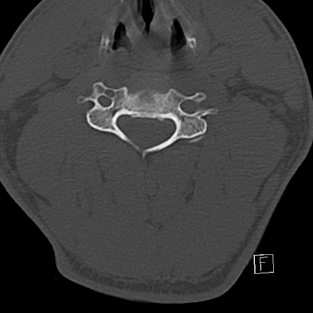
Gross anatomy
small, oval-shaped vertebral bodies
relatively wide vertebral arch with large vertebral foramen
relatively long, bifid (except for C7) inferiorly pointing spinous processes
transverse foramina protecting the vertebral arteries and veins
Osteology
Anterior components of the typical cervical vertebra 1:
body
posterolateral lip (uncus)
pedicle
-
transverse process
anterior and posterior tubercle of the transverse process
intertubercular lamella of the transverse process
foramen of the transverse process
Posterior components of the typical cervical vertebra 1:
lamina
bifid spinous process
superior articular process
inferior articular process
Articulations
intervertebral disc (superior and inferior): interposed between hyaline cartilage on the centrum of the vertebral bodies
uncovertebral joint 2: the superior surface of the vertebra below curves upward to form a hyaline covered lip. The lip articulates with the inferior bevelled surface of the vertebra above; this occurs bilaterally, and thus the intervertebral foramen in cervical vertebrae is bordered anteriorly by both the cervical vertebrae from above and below (as opposed to above alone)
-
facet (zygapophyseal) joint: articular processes lie at the junction of the pedicle and lamina, and the articular surface can be viewed as a cylinder sliced obliquely
upper facets face obliquely up and back
lower facets face down and forward
Blood supply
arterial: segmental branches from ascending cervical and vertebral arteries
venous: basivertebral veins, internal vertebral venous plexus, external vertebral venous plexus
Variant anatomy
variable presence of bifid spinous processes
variable length of spinous processes
blocked or fused vertebrae
accessory articulation between cervical transverse processes 4
Radiographic features
Plain radiograph
lateral view: if the patient is supine, this view will allow for all 7 vertebrae to be seen 1
swimmers view: another lateral view where the patient will have one arm up and one down 1; provides views of the cervicothoracic junction
AP view 1
AP open mouth: allows for assessment of C1 and C2 alignment and the dens
oblique view: for facet joints and intervertebral foramina 1


 Unable to process the form. Check for errors and try again.