
The Valsalva manoeuvre is the forced expiration of air against a closed airway, resulting in increased intra-abdominal, intrathoracic, and pharyngeal pressure. It can be performed against a closed glottis or by one closing the mouth and pinching the nose while forcibly exhaling.
It is commonly used to equalise the pressure in the middle ears (when changing altitude or diving underwater) and sinuses (if obstructed).
Terminology
The haemodynamic changes which occur during the manoeuvre may be divided into four phases; strain (phase I), hypotension (II), release (III), and pressure overshoot (IV). Relevant haemodynamic features include the following:
-
phase I
begin of strain, increase in systolic blood pressure (SBP) from increased transmural pressure across thoracic aorta
-
phase II
systolic BP falls secondary to the intrathoracic pressure decreasing venous return, and consequently, cardiac output
baroreceptor mediated elevation in heart rate and systemic vascular resistance
-
phase III
glottis is opened and the intrathoracic pressure normalises
systolic BP decreases
-
phase IV
restoration of cardiac output against an elevated SVR leads to an increased BP (MAP = CO x SVR)
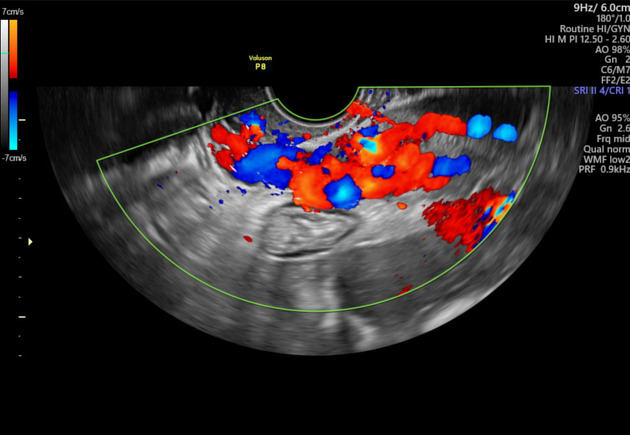
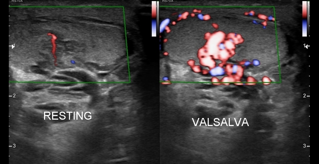
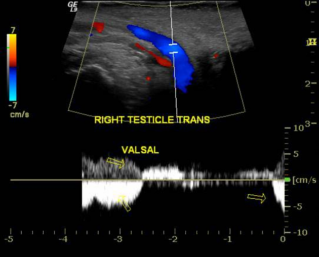
It is incorporated in medical imaging for dynamic assessment of various diseases such as:
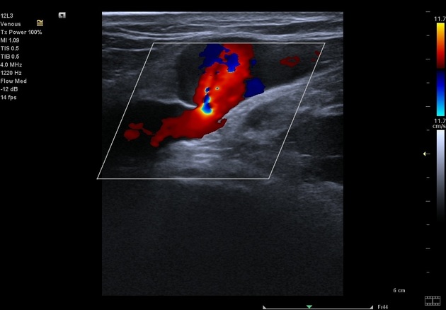
head and neck venous malformations
oesophageal reflux and hiatus hernias
leg deep venous thrombosis, chronic venous insufficiency and varicose veins
pelvic congestion syndrome: varicocele of the ovarian veins
The manoeuvre is employed in CT and MRI scanning of the neck to distend the pharynx for mucosal assessment. It is also a known cause of poor vascular contrast opacification causing transient contrast bolus interruption (termed transient interruption of contrast) 3, particularly in CTPA to investigate pulmonary embolism.
The manoeuvre is also a known cause of pneumomediastinum.
History and etymology
Antonio Maria Valsalva (1666-1723) was an Italian anatomist 1 who studied the anatomy of the ear and described the manoeuvre as a method of testing the patency of the Eustachian tube and also expelling pus from the middle ear.


 Unable to process the form. Check for errors and try again.