
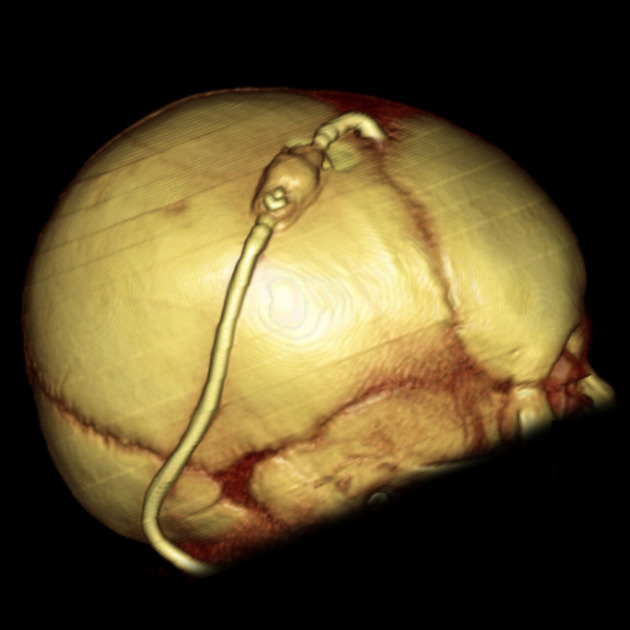
Ventriculoperitoneal (VP) shunts are devices used to shunt cerebrospinal fluid in the treatment of hydrocephalus.
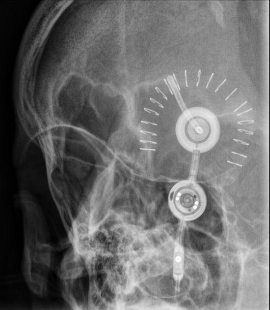
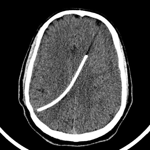
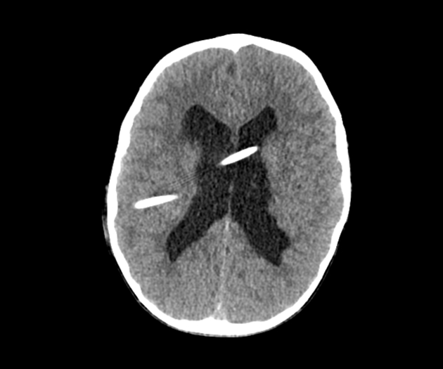
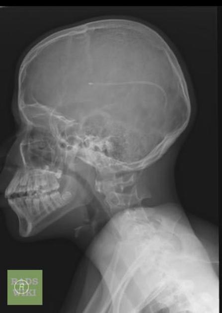
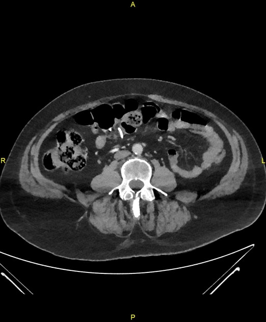
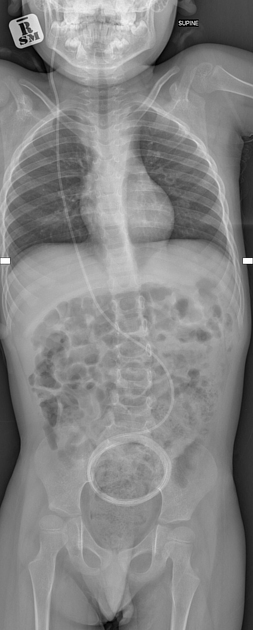
A catheter is placed with its tip in the ventricle. The external portion of the catheter is connected to a valve that regulates the flow of CSF based on pressure. This is commonly adjustable to allow regulation of intracranial pressure adjustments. Valves can have a gravitational anti-siphon unit. The distal catheter is tunnelled under the skin and into the peritoneal cavity.
Several other similar devices can be involved in the shunting of fluid from one cavity under pressure to another cavity of lower pressure:
ventriculoatrial shunt (CSF shunted into the vascular system)
lumboperitoneal shunt (CSF shunted from the spine)
cystoperitoneal shunt (cyst contents shunted to peritoneal space)
programmable shunt with variable pressing settings
On this page:
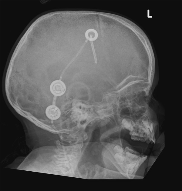
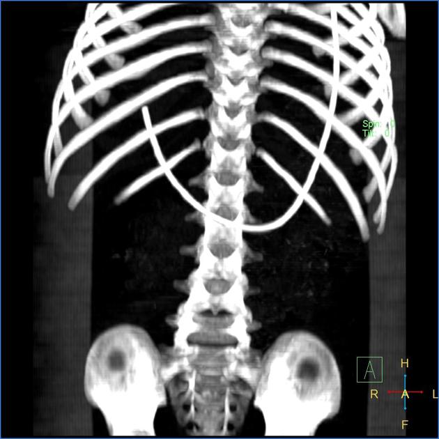
Radiographic features
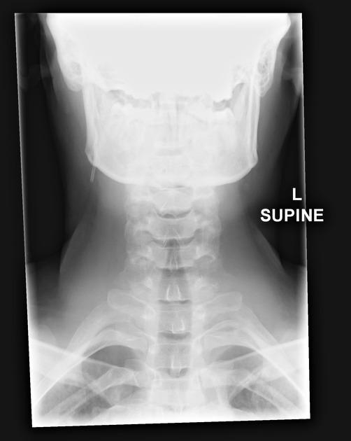
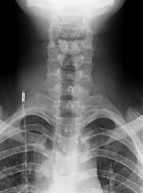
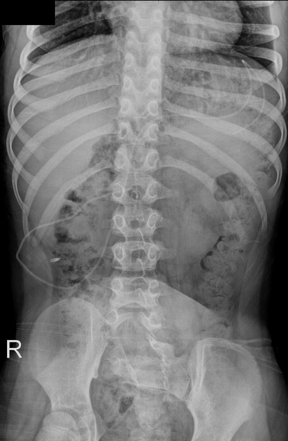
A shunt series is performed when there is concern about the normal functioning of a VP shunt.
Complications
Recognised complications include 1,2:
epidural haematoma, ipsilateral or contralateral (rare)
-
infection
-
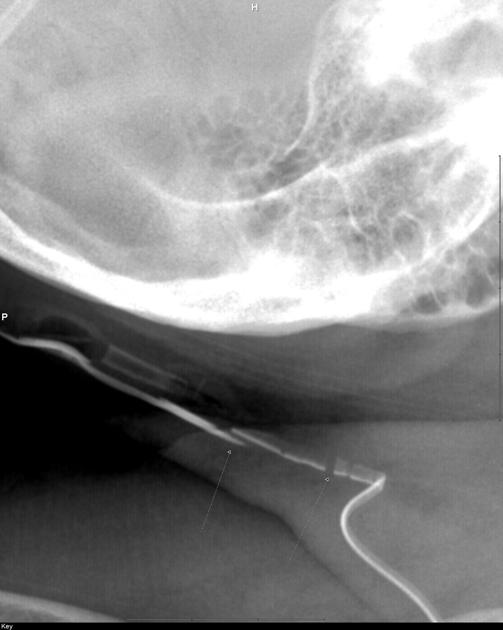
shunt malfunction
disconnections/breaks (most common in the neck)
migration
leakage
-
shunt overdrainage
intracranial peri-shunt fluid collection with oedema 3
trapped ventricle: after lateral ventricular shunting
-
distal complications
rarely, the distal end can encircle the bowel and cause strangulation 4


 Unable to process the form. Check for errors and try again.