
White matter buckling sign
Last revised by Karwan T. Khoshnaw
on 16 Nov 2018
Citation, DOI, disclosures and article data
Citation:
Gaillard F, Khoshnaw K, Hacking C, et al. White matter buckling sign. Reference article, Radiopaedia.org (Accessed on 28 Mar 2025) https://doi.org/10.53347/rID-36988
rID:
36988
Article created:
Disclosures:
At the time the article was created Frank Gaillard had no recorded disclosures.
View Frank Gaillard's current disclosures
Last revised:
16 Nov 2018,
Karwan T. Khoshnaw
Disclosures:
At the time the article was last revised Karwan T. Khoshnaw had no recorded disclosures.
View Karwan T. Khoshnaw's current disclosures
Revisions:
10 times, by
7 contributors -
see full revision history and disclosures
Systems:
Sections:
Tags:
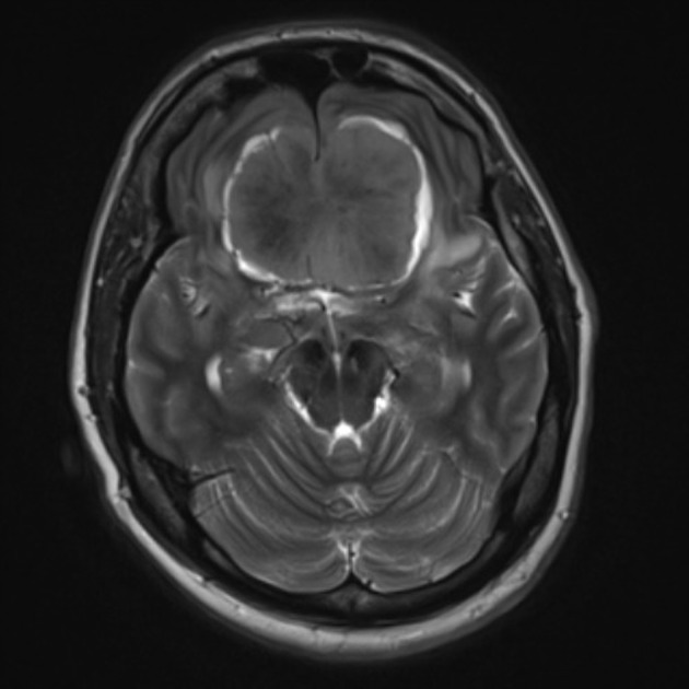
The white matter buckling sign is helpful in distinguishing an extra-axial intracranial mass from an intra-axial one and represents the white matter projecting into gyri being compressed and displaced by the mass, even in the presence of oedema (which would usually expand gyri, if the mass were intra-axial) 1.
References
- 1. George AE, Russell EJ, Kricheff II. White matter buckling: CT sign of extraaxial intracranial mass. AJR Am J Roentgenol. 1980;135 (5): 1031-6. doi:10.2214/ajr.135.5.1031 - Pubmed citation
Incoming Links
Cases:
- Parafalcine meningioma with CSF cleft sign
- Petroclival meningioma
- Meningioma - supratentorial
- Giant intracranial aneurysm
- Planum sphenoidale meningioma
- Cystic meningioma
- Meningioma
- Double meningioma
- Olfactory groove meningioma
- Spoke-wheel sign - meningioma
- Meningioma
- Olfactory groove meningioma
- Olfactory groove meningioma
- Planum sphenoidale meningioma
- Traumatic brain injury with epidural haematomas and skull fracture
- Traumatic brain injury with epidural haematomas and skull fracture


 Unable to process the form. Check for errors and try again.