
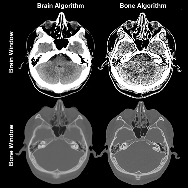
Windowing, also known as grey-level mapping, contrast stretching, histogram modification or contrast enhancement is the process in which the CT image greyscale component of an image is manipulated via the CT numbers; doing this will change the appearance of the picture to highlight particular structures. The brightness of the image is adjusted via the window level. The contrast is adjusted via the window width.
On this page:
Window parameters
Window width
As the name suggests, the window width (WW) measures the range of CT numbers in an image.
Therefore, a wider window width (2000 HU) will display a wider range of CT numbers. Consequently, the transition of dark to light structures will occur over a larger transition area to that of a narrow window width (<1000 HU).
Accordingly, it is important to note, that a significantly wide window displaying all the CT numbers will result in different attenuations between soft tissues to become obscured 1.
Wide window
Defined as 400-2000 HU best used in areas of acute differing attenuation values, a good example is lungs or cortical tissue, where air and vessels will sit side by side.
Narrow window
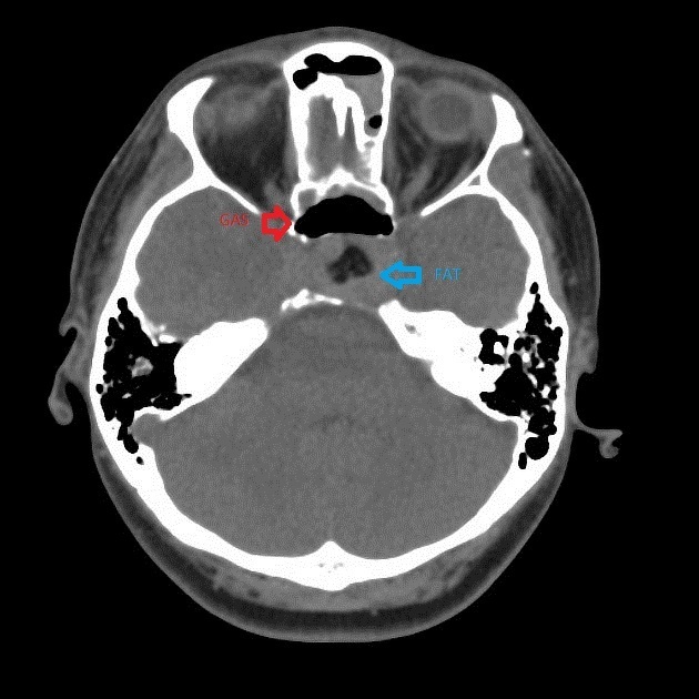
Defined as 50-350 HU are excellent when examining areas of similar attenuation, for example, soft tissue. It is widely used in differentiating subtle soft tissue attenuation differences i.e stroke imaging 5.
Window level/center
The window level (WL), often also referred to as the window center (WC), is the midpoint of the range of the CT numbers displayed.
When the window level is decreased the CT image will be brighter and vice versa.
Upper and lower grey level calculation
When presented with a WW and WL one can calculate the upper and lower grey levels i.e. values over x will be white and values below y will be black.
the upper grey level (x) is calculated via WL + (WW ÷ 2)
the lower grey level (y) is calculated via WL - (WW ÷ 2)
For example, a brain is W:80 L:40, therefore, all values above +80 will be white and all values below 0 are black.
Typical window width and level values
Although this varies somewhat from institution to institution and vendor to vendor, window width and centers are generally fairly similar. The values below are written as width and level (W:x L:y) in Hounsfield units (HU).
-
head and neck
brain W:80 L:40
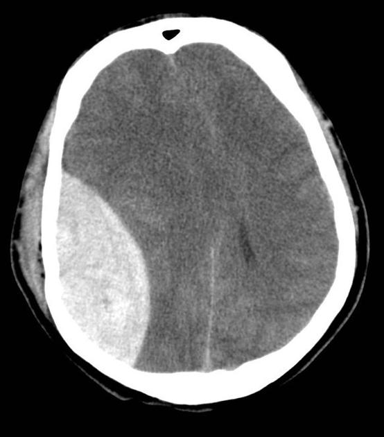
subdural W:130-300 L:50-100
stroke W:8 L:32 or W:40 L:40 3
temporal bones W:2800 L:600 or W:4000 L:700
soft tissues: W:350–400 L:20–60 4
-
chest
lungs W:1500 L:-600
mediastinum W:350 L:50
vascular/heart W: 600 L: 200 or e.g. W: 1000 L: 400
-
abdomen
soft tissues W:400 L:50
liver W:150 L:30
-
spine
soft tissues W:250 L:50
bone W:1800 L:400
Practical points
In certain cases, windowing is vital for imaging as pathology is better seen with a certain adjusted window i.e. lung window for HRCT Thorax.


 Unable to process the form. Check for errors and try again.