X-ray
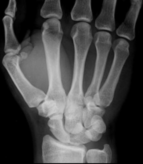
Note the triangular shape of the lunate on the PA projection. The lateral view shows volar dislocation of the lunate. The remaining carpal bones are in normal alignment.
 Frontal
 Oblique