CT
- CT abdomen and pelvis
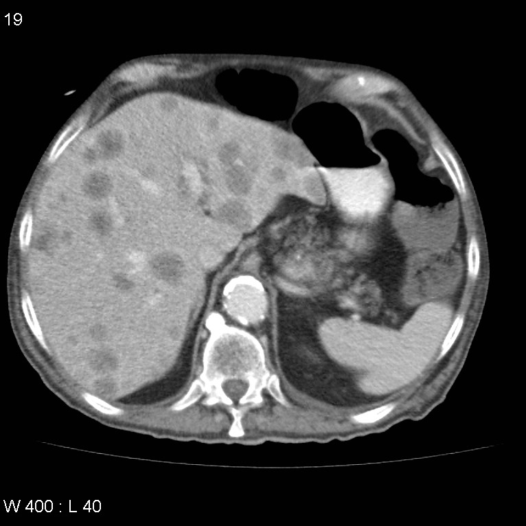
Axial C+ portal venous phase
Multiple abnormalities are present.
There is a diffuse abnormality of the distal colon, sigmoid colon and rectum, characterised by bowel wall thickening and peri-colonic fat stranding. This occurs in the region of presumed radiotherapy treatment and would be consistent, in this setting, with radiation colitis. The more proximal large and small bowel loops are prominent suggesting a degree of functional obstruction.
Multiple liver lesions with low attenuation and ill-defined margins. Similarly, multiple pulmonary lesions consistent with multiple metastases.
Large retroperitoneal lipoma measuring approximately 11cm in diameter, with vessels seen to coarse through this region.
The right adrenal gland is bulky with either an adenoma or a further metastasis. The pancreas and kidneys are normal with small cortical cysts seen within the kidneys but no evidence of hydronephrosis. Bilateral fat-containing inguinal hernias, with multiple prominent lymph nodes demonstrated within the left inguinal region, the largest of which has a short-axis diameter of 1.5cm.
Review of the bones demonstrates mixed sclerotic and lucent lesion involving most of the left hemipelvis consistent with malignant disease. A further soft tissue deposit is demonstrated within the coccyx/distal sacrum measuring approximately 4.2 x 2.8cm. Subtle sclerosis in the L4 vertebral body extending into the posterior elements are suspicious for further metastatic disease. An IDC is demonstrated within a contracted bladder which has some mild thickening and some hyperdensity in the dependent portions which may represent radiation cystitis and clinical correlation is recommended.