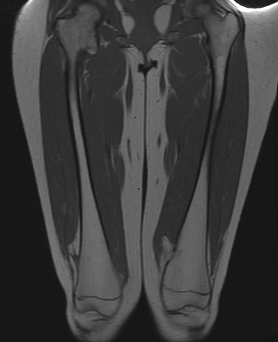
Coronal T1
 Coronal T1
 Axial T2
 Axial T2 fat sat
After a plain radiograph, MRI was performed.
Multiple
exostoses of the proximal and distal femora, as well as the proximal tibiae and
fibulae. The largest left sided exostosis is seen posteromedially
in the distal femoral metaphysis in the coronal and axial T1W MRI images. The hamstring and pes
anserine muscles and popliteal neurovascular structures are displaced by this
large exostosis. The cartilaginous cap measures 4mm thick, as seen in the axial T2W
MRI image.