
Left ventricular pseudoaneurysm and ventricular septal defect following left circumflex distribution myocardial infarction
Presentation
Three days of chest pain and shortness of breath.
Patient Data
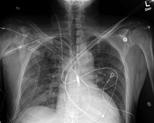
Impella device and pulmonary artery catheter appropriately positioned, no pneumothorax. Small posteriorly layering right pleural effusion, mild pulmonary edema.
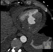

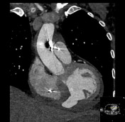
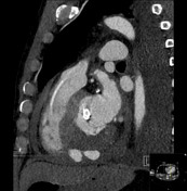
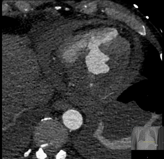
Decreased perfusion of the inferior and inferior septal walls. Defect inferior and lateral left ventricular wall with adjacent irregular outpouching indicating pseudoaneurysm. Muscular ventricular septal defect involving the inferior septum. Small pericardial effusion with increased density fluid, likely hemopericardium.
The coronary arteries arise normally with left coronary artery dominance. There is a stent in the left circumflex artery stent, with moderate LAD and left circumflex artery calcifications.
Bilateral moderate pleural effusions.
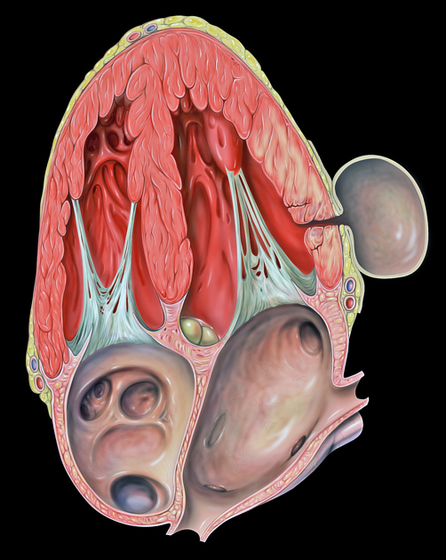
The illustration shows the pathophysiology of cardiac pseudoaneurysms: myocardial rupture contained by pericardium or other surrounding tissue.
Image credit: Patrick J. Lynch (medical illustrator)
No changes were made to the original image.
For full attribution, see case discussion.
Comment: Patrick J. Lynch; illustrator; C. Carl Jaffe; MD; cardiologist Yale University Center for Advanced Instructional Media Medical Illustrations by Patrick Lynch, generated for multimedia teaching projects by the Yale University School of Medicine, Center for Advanced Instructional Media, 1987-2000.
Case Discussion
Pseudoaneurysms and ventricular septal defects uncommonly occur after myocardial infarction. This patient was unfortunate and had both, but was treated and survived. The involvement of both the lateral ventricular wall (pseudoaneurysm) and the inferior septum (VSD) is likely a consequence of ischemia in the distribution of a dominant circumflex coronary.
Image attribution: pseudoaneurysm of the left ventricle - by Patrick J. Lynch
Author: Patrick J. Lynch (medical illustrator)
Original file: https://commons.wikimedia.org/wiki/File:Heart_pseudoaneurysm_a4c.jpg
Modification: no changes were made to the original image


 Unable to process the form. Check for errors and try again.