
Presentation
Nasal obstruction and epistaxis with endoscopic findings of a mass in the nasopharynx.
Patient Data


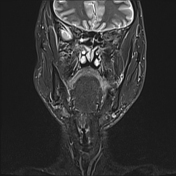
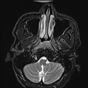

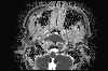
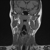
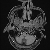
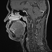
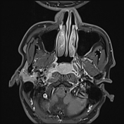
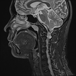
A nasopharyngeal mass measuring 15 x 26 x 27 mm exhibits high signal intensity on STIR and T2FS sequences, intermediate signal intensity on T1W images, restricted diffusion (bright on DWI and dark on ADC), and strong homogeneous enhancement after contrast administration. The lesion appears localized with no evidence of surrounding invasion.
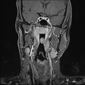
Several highly suspicious lymph nodes are identified in the right parapharyngeal space and right cervical region near the right parotid gland, with a maximum diameter of 19 x 14 mm. These nodes are round, exhibit irregular margins, restricted diffusion, and demonstrate strong homogeneous enhancement on the post-contrast study.

Macroscopic:
The specimen consists of two fragments, measuring 0.5 cm each, with a light brown color.
Microscopic:
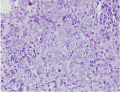
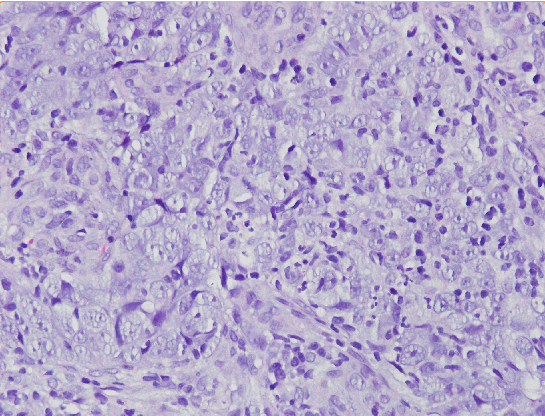
Clusters of poorly differentiated epithelial cells are observed invading a necrotic inflammatory fibrous stroma. The nuclei exhibit hyperchromasia and pleomorphism, with a high mitotic rate.
Conclusion:
Poorly differentiated carcinoma.
Recommendation:
Immunohistochemical staining for P16 and P40 is suggested.
Case Discussion
The imaging and histopathological findings are consistent with nasopharyngeal carcinoma, which can be classified as T1 N1 M0 according to the AJCC 2018 staging system.
Unfortunately, for some personal reason, the patient defaulted follow up and did not undergo further tests or receive treatment, so the final outcome could not be followed up.


 Unable to process the form. Check for errors and try again.