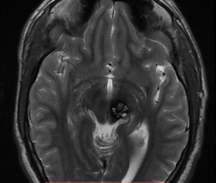
Axial T2
 Axial FLAIR
 Sagittal T1
A rounded lesion with peripheral signal drop-out consistent with hemosiderin is seen in left side of midbrain. Centrally the lesion is heterogeneous in signal with regions of high signal on both T1 and T2 weighted images. There is no peripheral oedema and no evidence of hydrocephalus.
Features are characteristic of a midbrain cavernous malformation.