Coronal C+ arterial phase
Sagittal C+ arterial phase
The 30 x 40 mm mass located above the right petrous apex, involving Meckel's cave, is inseparable from the postero-lateral margin of the clivus, and has coarse central calcifications. There is local destruction of cortex.
Note that the right supraclinoid internal carotid artery has a close relationship to the mass, and that the right posterior cerebral artery is almost entirely fetal origin, the branch displaced by and covered by the mass.
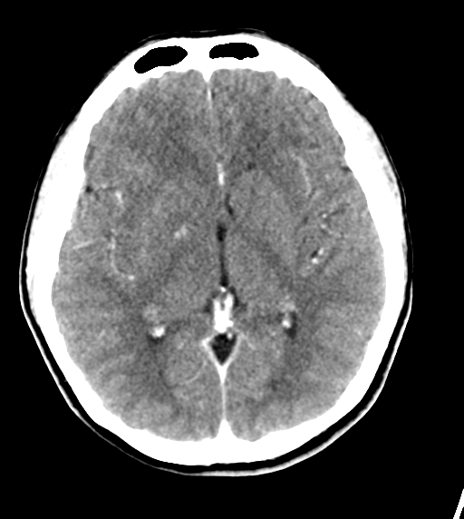
Extracranial vessels are normal. No significant stenosis. No intracranial aneurysm or vascular malformation.
Conclusion
The additional information from the CT study effectively excludes neurofibroma or meningioma. Either chondrosarcoma or chordoma are the most likely diagnoses-the T2 signal characteristics more in favour of a chondrosarcoma. Note the close relation to the fetal right posterior cerebral artery which is displaced and on the medial border of the tumour, between right cerebral peduncle and postero-medial tumour margin.