Ultrasound
- First trimester scan
Thick tubular left adnexal structure inbetween the left uterine cornu and left ovary reaching 2 cm in thickness with hypoechoic heterogeneous content, likely representing left Fallopian tube with edematous mucosa and retained hemorrhagic content. It shows abrupt rounded expansion at the isthmus part measuring 2 x 2.5 cm with internal cystic structure measuring 1 x 1.5 cm surrounded by echogenic periphery (decidual reaction) showing a Yolk sac with no detected fetal pole or internal vascularity at very low Doppler settings. Features raise the possibility of heterotopic pregnancy of uncertain viability.
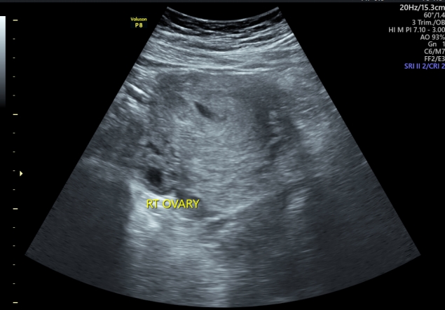
Normal both ovaries. No free fluid at the abdomen or pelvis.
A single viable intrauterine fetus is noted, with active fetal movements and visible cardiac pulsations. (FHR= 147 bpm). Normal biophysical profile. The placenta is located anteriorly with grade zero maturation. Intact placental bed. Apart from a heterogeneous hypoechoic area near the left cornu measuring 2.2 x 1.8 cm, suggestive of organized retroplacental hemorrhagic extension from the left Fallopian tube disturbed ectopic pregnancy.