
Presentation
Right frontal non-painful hard swelling.
Patient Data


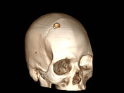
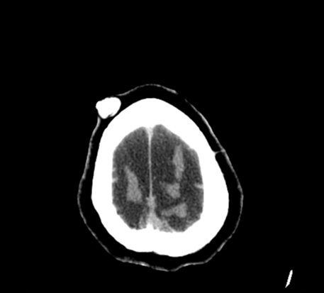
A well-defined partially calcified ovoid scalp mass is seen at the right frontal region (corresponding to the clinically palpable swelling). Other smaller similar lesions are seen at the right temporal region.
Cavum septum pellucidum & verge (variant).
Involutional brain changes are also noted & manifested by prominent ventricular system, subarachnoid CSF spaces, cortical sulci, basal cisterns and Sylvian fissures.
Case Discussion
CT findings are in keeping with proliferating trichilemmal cysts (also known as pilar cysts).
Proliferating trichilemmal cysts usually arise in the scalp. They have a female predominance.
An epidermal inclusion cyst is the most common lesion to be considered in the differential diagnosis.


 Unable to process the form. Check for errors and try again.