
Presentation
Chronic headaches with left hearing loss.
Patient Data
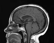

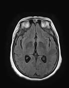
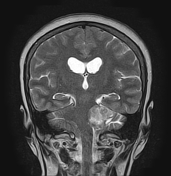
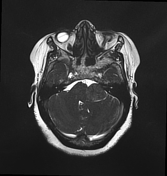
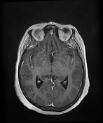

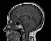
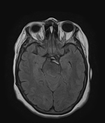
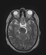
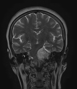
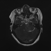
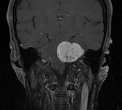
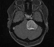
The MRI sequences demonstrate a solid extra-axial mass at the left cerebellopontine angle measuring (28 x 24 x 22 mm) with mild enlargement of the ipsilateral porus acusticus. It displays a low signal intensity on T1WI, heterogeneous high signal intensity on T2WI/FLAIR with heterogeneous enhancement and central necrosis on postcontrast sequences.
A mass effect is noted on the brainstem, left middle cerebellar peduncle, a cisternal portion of the trigeminal nerve as well as the cerebellar hemisphere and 4th ventricle which is laminated and displaced to the right
Moderate dilatation of the 3rd and lateral ventricles, indicating obstructive hydrocephalus.

The patient refused surgery. A VP shunt was inserted and an MRI exam was performed 15 months later showing an increase in the size of the left cerebellopontine mass measuring (42 x 36 x 34 mm) as well as an increase of the mass effect on the adjacent structures with mild ipsilateral cerebellar oedema.
Case Discussion
MRI features of a left acoustic neuroma, compressing the adjacent structures with obstructive hydrocephalus treated by VP shunt.


 Unable to process the form. Check for errors and try again.