
Presentation
Direct trauma to the top of the right shoulder.
Patient Data
Age: 30 years
Gender: Female
From the case:
Acromial fracture


Download
Info
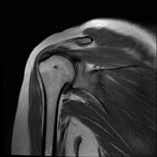
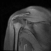
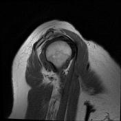
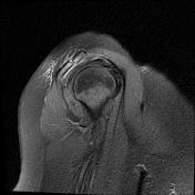
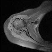
Mildly displaced vertical fracture line with surrounding bone marrow oedema/bruise is seen through the acromial process middle third portion.
Low lying type II acromion also is noted.
The acromioclavicular joint is unremarkable.
Subacromial/subdeltoid bursal effusion is present.
Abnormal intrasubstance increased fluid signal and thickening is present along with the supraspinatus tendon fibres related to tendinosis.
The bifid biceps tendon is in the bicipital groove and has a normal appearance.
Case Discussion
Isolated acromial fracture without associated shoulder injuries rarely occurs and often associated with rotator cuff injury and impairment of shoulder function.


 Unable to process the form. Check for errors and try again.