
Presentation
Large right suprarenal lesion found incidentally on presenting CT (not shown). CT with contrast to evaluate further.
Patient Data
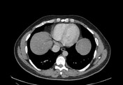

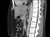
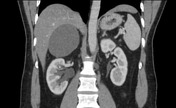

8 x 8 cm cystic lesion (axial, coronal) with partial mural calcification inseparable from the right adrenal gland and in close proximity to the superior pole of the right kidney. 2.5 x 1.5 cm lobulated component arises from the inferolateral aspect of the lesion.
Right-sided moderate hydronephroureterosis (secondary to 5 mm calculus in the distal
ureter - off image). 4 mm non-obstructing calculus in the right upper pole and 5 mm
non-obstructing calculus in the right interpolar region. A couple of tiny non-obstructing calculi in the left upper renal pole.
Liver, gallbladder, spleen, pancreas and left adrenal unremarkable.
No ascites. No nodal enlargement. No bowel obstruction.
Conclusion:
1. Large cystic lesion likely right adrenal origin
2. Obstructed right kidney
Case Discussion
An ultrasound performed eight years earlier (images not available) showed a similar slightly smaller cystic lesion in the right adrenal region.
Adrenal cysts are rare. Usually they are benign.
In 1966, Dale G Foster subclassified adrenal cysts into four main types based on histopathological evaluation of 115 cases: epithelial cysts, parasitic cysts, especially hydatid, pseudocysts and endothelial cysts. In this series, endothelial cysts were the commonest type. Although other studies since have found pseudocysts to be more common 2.
It was decided that since the cyst in this case was an incidental pick up and the patient was not symptomatic from it to manage it conservatively. Therefore the exact type of cyst has never been ascertained.


 Unable to process the form. Check for errors and try again.