
Presentation
Presenting to the emergency department with headache, dizziness, and left-side weakness.
Patient Data


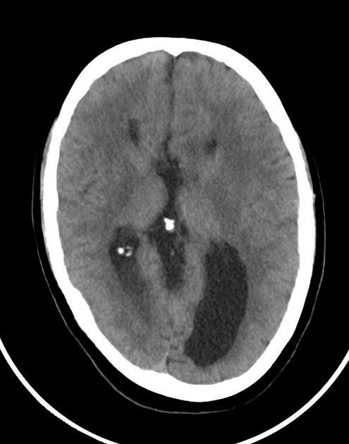
Agenesis of corpus callosum with colpocephaly and high riding third ventricle.
Small nodules isodense to gray matter at the lateral walls of both lateral ventricles suggesting subependymal heterotopia.
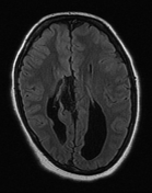
Right parasagittal interhemispheric cyst.
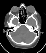
Extra-axial cystic lesion at the right side of the posterior cranial fossa causing mass effect on the right cerebellar hemisphere suggesting arachnoid cyst.
Fusiform focal lesion centered on the right optic nerve.

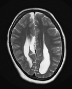
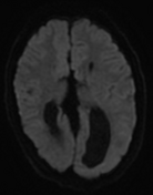
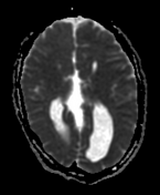
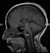
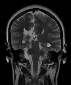
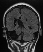
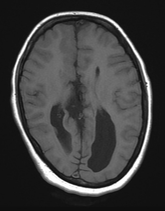
Agenesis of the corpus callosum with widely separated parallel bodies of lateral ventricles, colpocephaly, high riding third ventricle communicating with interhemispheric cistern, Absent cingulate gyrus with radial medial hemispheric gyri on sagittal view, and moose head appearance of lateral ventricles on coronal view.
Right medial frontal polymicrogyria.
Right parasagittal interhemispheric cyst.
Cystic lesion at the right aspect of the posterior cranial fossa causing mass effect on the right cerebellar hemisphere suggesting arachnoid cyst.
Relatively small right eye globe (microphthalmos) with right optic nerve cystic lesion.
Case Discussion
This patient is a known case of Aicardi syndrome with typical radiological features.


 Unable to process the form. Check for errors and try again.