
Presentation
Patient presenting with enlarging left breast mass and left axillary adenopathy.
Patient Data
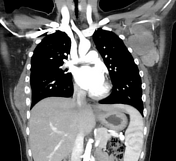

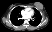

In the left axilla, there is a conglomerate of nodular lesions with heterogeneous enhancement, iso-to-hypoenhancing relative to adjacent muscle. There is a slightly separate nodular lesion in the surrounding soft tissues. The lesions extend to the left infraclavicular space with stranding of surrounding fat. In the topography of the left breast, there is a similar appearing conglomerate of multiple hypoechoic attenuating nodular lesions with secondary protrusion of the left breast contour.
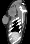
In the right lateral chest wall, there is a heterogeneously enhancing ovoid soft tissue mass, predominantly hypoattenuating and extending into the chest wall from the lateral right 4th-5th intercostal space. The mass extends inferiorly to the 7th-8th intercostal space with periosteal thickening and sclerosis suggesting bony invasion. The mass invades the adjacent chest wall into the intrathoracic structures with thickening of adjacent pleura.
Case Discussion
A core needle biopsy was performed.
Histology:
Gross: ten cores of pink-tan soft tissues measuring 1.5 to 2.2 cm from left breast mass. Ten cores of pink-tan soft tissues measuring 1.0 to 2.0 cm from left axillary mass.
Microscopy: soft tissues consist of neoplastic infiltrate. The neoplastic cells are pleomorphic with oval-shaped nuclei containing coarse chromatin and small nucleoli with a small amount of eosinophilic cytoplasm. There are areas of geographic tumour necrosis. There are scattered tingible body macrophages among tumour cells. Mitosis is brisk.
Immunohistochemical stains: positive for CD45, CD3, ALK-1, CD30, EMA (focal), CD99 (focal). Negative for CD20, myogenin, desmin, cytokeratin, Phox2B, NKX2.2, Bcl-2, PAX5, CD10. Ki-67 stains 70% of tumour cells. BCL6 stains nucleus of tumour cells.
Flow cytometry shows aberrant CD30-positive large cell population. FISH study shows positive ALK gene rearrangement.
Final diagnosis: ALK-positive anaplastic large cell lymphoma


 Unable to process the form. Check for errors and try again.