
Presentation
Progressive deterioration of vision of the right eye. The ophthalmologist suspected compressive optic nerve atrophy.
Patient Data
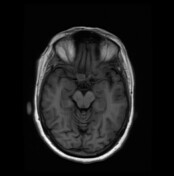

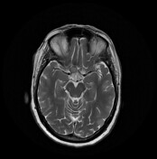
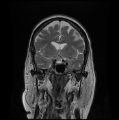
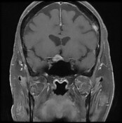
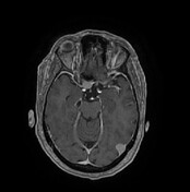
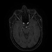

An ovoid lesion is seen within the right aspect of the suprasellar region/cistern. It is intimately related and inseparable from and even encases the posterior retrobulbar part of the right optic nerve as well as related part of the optic chiasm. It encroaches upon the related part of the suprasellar cistern. The lesion is seen in close relation and compresses the most distal part of the right internal carotid artery and right A1 segment. It appears isointense in T1 & T2 and shows avid homogenous enhancement in the post-contrast study with enhancing meningeal tail. It shows no remarkable restricted diffusion in DWI or blooming in gradient-echo sequence. Another similar lesion is seen in the left occipital extra-axial region.
Case Discussion
MRI features are suggestive of meningioma of the anterior clinoid process. The described site is not a typical location of meningioma. Meningioma is the most probable diagnosis.


 Unable to process the form. Check for errors and try again.