
Presentation
The patient presents with a congenital, anterior fontanelle cystic swelling.
Patient Data
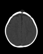

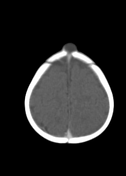
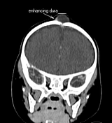
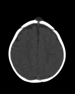
There is a well-defined cystic mass overlying the anterior fontanelle. It measures 15.0 x 12.0 x 13.6 mm (w x ap x cc ).The lesion is superficial to the dura, with no intradural or intracranial extension. There is no associated calcification and no abnormal enhancement. There is widening and remodelling of the underlying anterior fontanelle well demonstrated by the 3D reformats. Contrast-enhanced CT brain is otherwise unremarkable.
CT courtesy of Dr Pearl GNM Quvane.
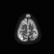
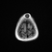
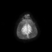
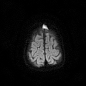
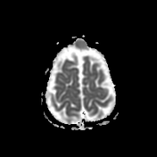
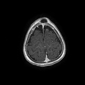

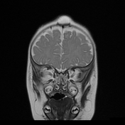
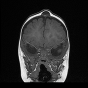
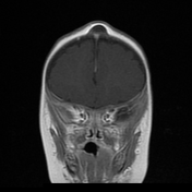
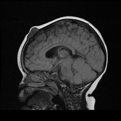
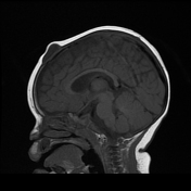
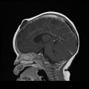

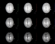
Multiparametric contrast-enhanced MRI brain confirms a CSF intensity (rather than fat intensity), non-enhancing, cystic mass lesion overlying the anterior fontanelle. There is restriction on diffusion imaging with reduced average diffusivity on ADC mapping suggesting a likely epidermoid cyst. The regional dura is well-identified and intact.
MRI courtesy of Dr IA. Nagdee.
Case Discussion
Anterior fontanelle inclusion cyst is a more appropriate term for these congenital dermoid/epidermoid cystic lesions that overlie the anterior fontanelle without any intracranial or intradural extension on imaging. In this instance, the lesion measured CSF intensity on the CT brain (-50 HU) and followed CSF intensity on MRI. There is however diffusion restriction and confirmed reduced average diffusivity on ADC mapping suggesting it to be an epidermoid cyst in nature.


 Unable to process the form. Check for errors and try again.