
Presentation
The patient presented to emergency department complaining of dizziness. No signs of neurological deficit.
Patient Data
Age: 45 years
Gender: Male
From the case:
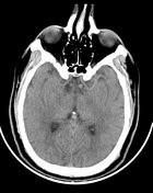
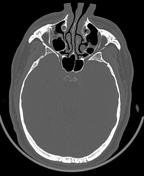
Anterior temporal encephalocele

Show annotations
Download
Info
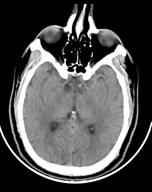
Anteroinferior protrusion of the anterior right temporal lobe with associated remodeling of the right greater wing of sphenoid (GWS) which appears thinned, anterior displaced with rarefaction.
Otherwise unremarkable CT brain.
From the case:
Anterior temporal encephalocele

Show annotations
Download
Info

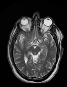
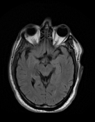
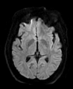
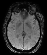
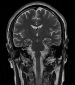
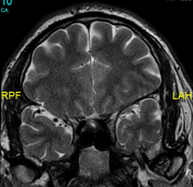
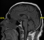
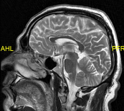
Anterior protrusion of anterior right temporal lobe through the greater wing of sphenoid (axial T2, coronal T2, sagittal T2).
Normal size and configuration of the ventricular system.
No midline shift.
No definite intra or extra-axial areas of abnormal intensity.
Within normal posterior fossa structures.
Case Discussion
The radiological features are characteristic of anterior temporal encephalocele.


 Unable to process the form. Check for errors and try again.