
Presentation
Partially imaged malacia at the T3 segment of the cord in the follow-up brain MRI of a stable clival mass.
Patient Data
Age: 70 years
Gender: Male
From the case:
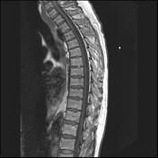
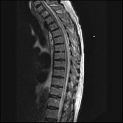
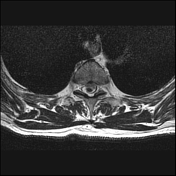
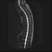
Anterior (ventral) cord herniation

Download
Info

There is focal anterior deviation of the spinal cord along the anterior dural margin at T3. There is some attenuation of the T2 void behind the spinal cord at the deviated and flat level in keeping with turbulent CSF flow. There is no significant decrease in volume cord, no myelopathy, no syringomyelia.
This finding might be related to an anterior dural adhesion or herniation of the cord through a small anterior dural defect.
Case Discussion
Case contributed by Dr Lorne Rosenbloom, neuroradiology assistant professor of McGill University.


 Unable to process the form. Check for errors and try again.