
Presentation
Right upper quadrant pain, fever and jaundice.
Patient Data


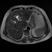
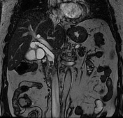
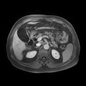
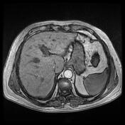
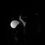
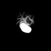
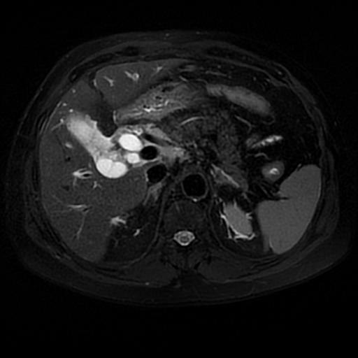
Distended gallbladder with homogeneous fluid content with no gallstones or significant wall thickening. Dilatation of the intrahepatic and common bile ducts (CBD = 19 mm) with choledocholithiasis (at least 5 stones). Wall thickening with enhancement mainly of the distal CBD. Periportal edema with small lymphadenopathy.
Case Discussion
The clinical presentation with Charcot triad (fever, right upper quadrant abdominal pain, and jaundice) and the MRI features are consistent with ascending cholangitis.
The diagnosis of ascending cholangitis (or acute cholangitis) is mainly clinical. The imaging is performed to determine if there is evidence of bile duct dilatation, bile duct wall thickening, cholelithiasis, or choledocholithiasis.


 Unable to process the form. Check for errors and try again.