
Presentation
Palpable high parietal subgaleal mass
Patient Data


Mid-line subcutaneous cystic lesion with vertically oriented primitive falcine vein pointing to the cephalocele.
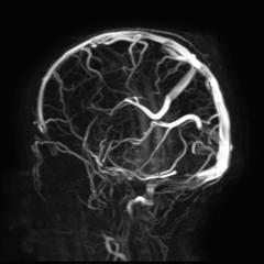
Vertically oriented persistent falcine vein.
Fenestrated superior sagittal sinus at the site of the cephalocele.
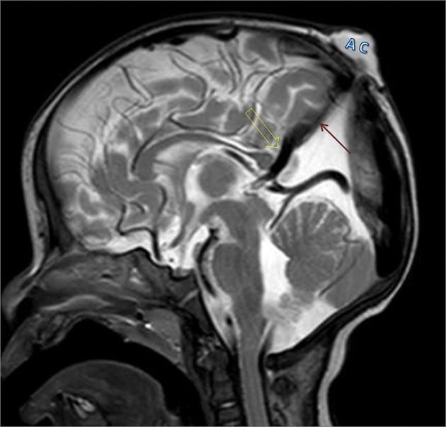
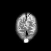
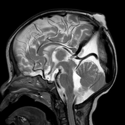
Sagittal T2 WI shows:
Mid-line subcutaneous cystic lesion (atertic cephalocele) (AC),
Vertically oriented persistent primitive falcine vein (open arrow) & an adjacent hardly distinct thin fibrous stalk (solid arrow) are pointing to and connecting the cephalocele through the calvarial defect.
Prominent superior cerebellar cistern & suprapineal recess.
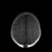
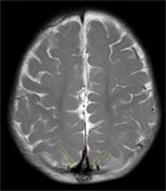
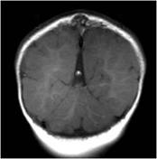
Axial T2 WI shows:
Fenestration of the superior sagittal sinus (open arrows) surrounding the cephalocele fibrous stalk (Solid arrow)
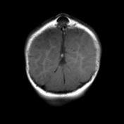
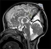
Coronal T1 WI shows:
Peaked configuration of the tentorium.
Case Discussion
Atretic cephalocele consists of:
- subgaleal mass with intracranial extension.
- vertical primitive falcine vein and fibrous tract point to the subcutaneous scalp mass.
- focal fenestration of the superior sagittal sinus at the AC.
Differential diagnosis:
- sinus pericranii
- dermoid or epidermoid sinus
- sebaceous cyst
- cephalohematoma
- infantile hemangioma


 Unable to process the form. Check for errors and try again.