
Presentation
Incidentally detected after a left knee injury due to a motorcycle accident.
Patient Data
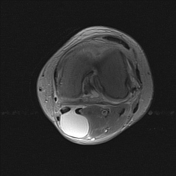

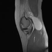
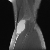
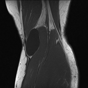

A fluid-filled cystic lesion is present in the medial popliteal fossa, measuring 26 × 24 × 50 mm. It is located between the medial head of the gastrocnemius tendon and the semimembranosus tendon, communicating with the knee joint through a narrow connection. The lesion has well-defined margins.
The patella appears elevated and shows signs of patellofemoral subluxation.
There is joint effusion, primarily involving the suprapatellar bursa.
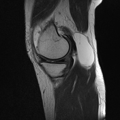
There are osteophytes at the margins of the femoral condyles and tibial plateau, accompanied by subchondral bone cysts in the bilateral tibial plateaus, suggesting osteoarthritis.
Grade III chondromalacia patellae (according to Outerbridge classification) at the posterior aspect of the patella.
Grade IV chondromalacia patellae (according to Outerbridge classification) at the medial aspect of the patella.
Case Discussion
The imaging findings are consistent with a popliteal synovial cyst (Baker's cyst) associated with knee osteoarthritis.
This is an incidental finding, with no clinical symptoms or complications (such as rupture, dissection, or compression) observed on imaging; therefore, no additional treatment is required.
If treatment is necessary, procedures such as aspiration, corticosteroid injection, and surgery may be considered to reduce the size of the Baker’s cyst and alleviate symptoms.


 Unable to process the form. Check for errors and try again.