
Presentation
The ulcerative lesion in the right-side orbital lateral canthus for five months.
Patient Data


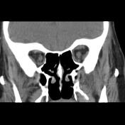
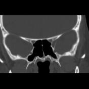
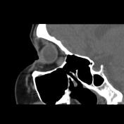
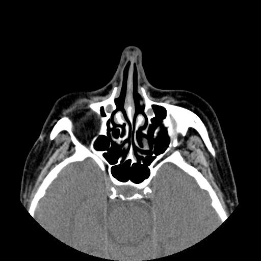
Circumferential Irregular ulcerative margin soft tissue thickening with thickness up to 8 mm in right orbital lateral canthus extended within related upper and lower eyelids, orbital septum, and lateral rectus muscle tendon insertion, lacrimal gland palpebral segment, and partially encased related eye uveo-scleral layer. Right eye enophthalmos, exodeviation, and no native lens are seen.
Case Discussion
The case illustrates the non-contrast MDCT features of the pathology-proved basal cell carcinoma nodular type of the lateral canthus. The most common presentation of the basal cell carcinoma is an ulcerative skin lesion, the tumour has a high recurrence rate, and the main treatment of the tumour is complete surgical excision. Canthal basal cell carcinoma has a higher risk of orbital invasion 1.


 Unable to process the form. Check for errors and try again.