
Presentation
Bilateral cervical painless swelling in a patient known for Graves disease.
Patient Data
Age: 50 years
Gender: Female
From the case:
Bilateral carotid body tumour


Download
Info
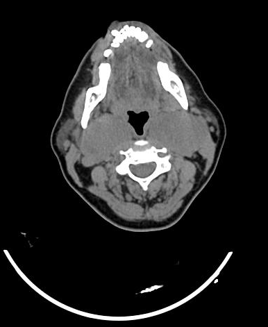
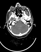
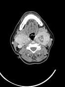
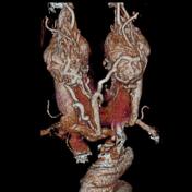
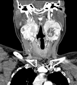
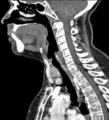
Bilateral well-defined upper cervical soft tissue mass, isodense to the muscles with vivid enhancement on postcontrast images, splaying the internal and external carotid arteries (lyre sign).
Diffusely enlarged thyroid gland with heterogeneous enhancement (known Graves disease).
Case Discussion
CT appearance of carotid body tumour (also called a chemodectoma or carotid body paraganglioma).
Carotid body tumours are the most common type of paraganglioma of the head and neck (account for 60-70%) and are bilateral in approximately 10% of cases (as in this case).


 Unable to process the form. Check for errors and try again.