
Presentation
Left upper limb weakness. Status post cervical disc surgery about 6 months ago.
Patient Data

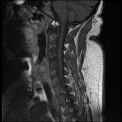
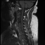
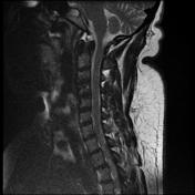

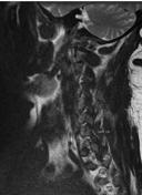
Evidence of C3/C4 and C4/C5 discectomy and cage placement.
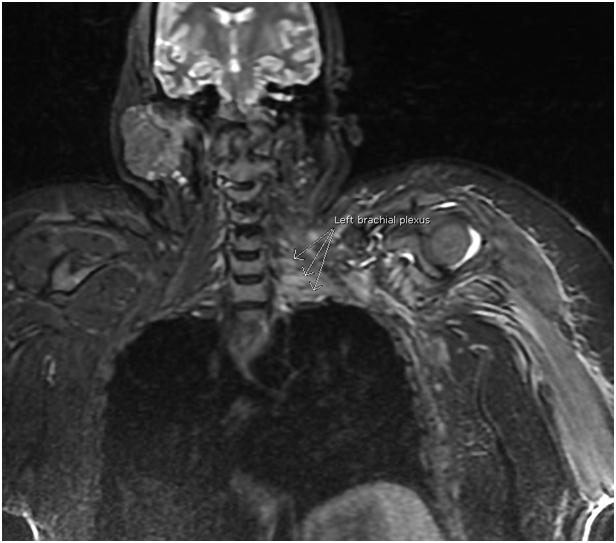
The lower cervical spinal nerves of the left brachial plexus, and their dorsal root ganglia from C5 to D1 are seen thickened eliciting slightly high T2/FLAIR signal with subtle post-contrast enhancement. No definite loss of fibre continuity or related masses.
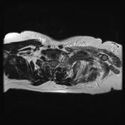
The left shoulder girdle muscles as well as left arm, pectoralis major and latissimus dorsi muscles show reduced girth with high T2/STIR signals and post-contrast enhancement.

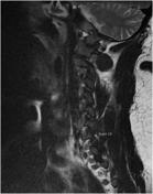
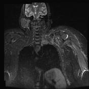
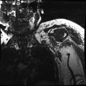
High FLAIR signal and swelling of the left brachial plexus.
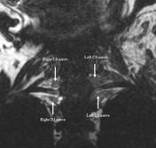
Large left brachial plexus roots, best noted at C8.
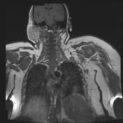
Normal right brachial plexus roots as seen here for comparison.
Coronal FIESTA explicitly shows the difference in size between left and right brachial plexuses.
Case Discussion
Left brachial plexopathy, likely postoperative sequelae due to excessive traction or malpositioning, or less commonly postoperative infection, with subsequent left shoulder girdle and left upper limb muscles atrophy and oedema.


 Unable to process the form. Check for errors and try again.