
Presentation
Slowly growing painless mass at the right buccal region over six months.
Patient Data


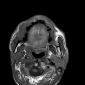
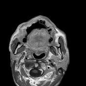
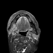
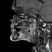
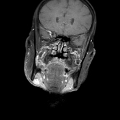
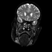
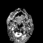
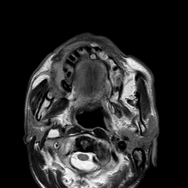
A well-defined heterogeneously enhancing right buccal mucosal lesion, measuring 3.2 x 1.7 x 3.7 cm. It displays high signal intensity (SI) on T2, low SI on T1, and restricted DWI, with ADC 0.9 x 10 -3 mm2. It is not separated from the superior and inferior gingivobuccal sulcus, contacting the superior and inferior alveolar margins, with subtle erosion of the superior alveolar cortex.
The mass infiltrates the right parotid duct (Stensen duct) with consequent small-sized (atrophic) right parotid gland.
Several enlarged right cervical lymph nodes at levels Ib and IIa with thickened cortex, largest measures 1.3 x 2.2 cm.
Case Discussion
Pathologically proven right buccal mucosal space squamous cell carcinoma, underwent wide local excision with supraclavicular artery island flap (SCAIF) and right supraomohyoid block neck dissection.
Histological examination:
Papillomatous tumour proliferation lined by hyperplastic atypical stratified squamous epithelial cells with dysplastic features, hyperkeratosis, and heavy lymphoplasmacytic infiltrate at the base.
Diagnosis: Verrucous carcinoma.
Free all surgical margins all around.
Negative for lymphovascular emboli and perineural invasion.
Free salivary gland tissue and 23 lymph nodes.


 Unable to process the form. Check for errors and try again.