
Presentation
The patient had twisted knee, felt pop with immediate swelling knee.
Patient Data

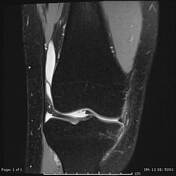
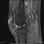

There is ACL full thickness tear.
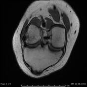
There is a displaced vertical meniscal tear with truncation and a small size of the body and the posterior horn of the medial meniscus. There is migration of the displaced fragment in the intercondylar notch (best seen in coronal sequences) with development double delta sign. Finding consistent with Displaced Bucket Handle tear.
There is sub-chondral oedema at the sulcus terminalis and the posterior aspect of the tibia in keeping with a pivot-shift injury but elsewhere the marrow returns a normal signal Patella is normal in position. There are few partial thickness cartilage fissuring seen within lateral patellar facet. Elsewhere cartilage is intact.
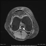
Moderate joint effusion. Tiny Baker cyst.
Case Discussion
The mechanical traumatic history together with radiological finding, all compatible with A case of medial meniscal bucket handle tear with other association like ACL tear with marrow oedematous changes


 Unable to process the form. Check for errors and try again.