
Presentation
Two-day history of visual abnormalities and gait disturbance.
Patient Data


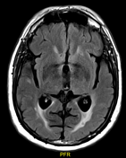
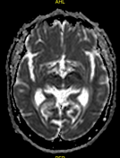
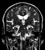
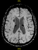
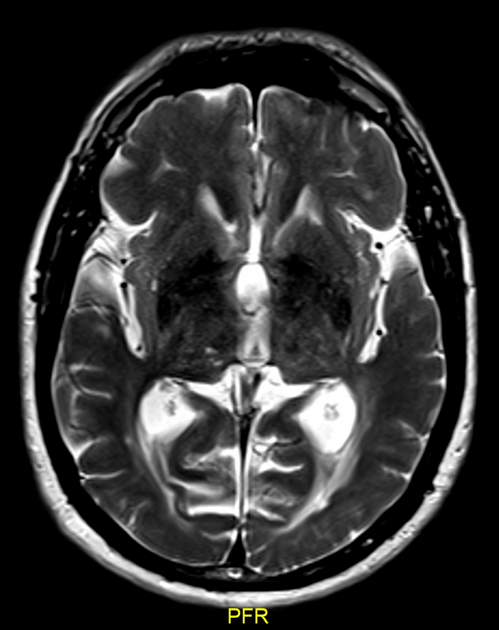
Bilaterally widespread cerebral, subcortical, and periventricular confluent white matter hyperintensities are easily appreciated on FLAIR and T2. These abnormalities are sparing the subcortical U-fibres and involve the temporal lobes and brainstem.
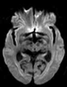
An abnormal focus of diffusion restriction is seen at the dorsal pons on the right side at the medial longitudinal fasciculus (MLF) site. It shows a low signal on ADC and a high signal on T2.
A few foci of microbleeds are seen on SWI.
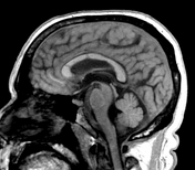
Chronic ischaemic changes are seen in the form of areas of encephalomalacia, with associated early-for-age involution changes in the form of deepened sulci and dilated ventricles as well as relative volume loss.
Case Discussion
The described brain changes, especially the temporal lobe white matter involvement and the sparing of the subcortical U-fibres, are consistent with cerebral autosomal dominant arteriopathy with subcortical infarcts and leukoencephalopathy (CADASIL).
The infarction seen at the medial longitudinal fasciculus (which coordinates movements of the eyes with movements of the head) can explain the patient's presentation.
Case courtesy: Prof. Dr. Yasser Ragab


 Unable to process the form. Check for errors and try again.