
Presentation
Sudden blurred vision in the right eye and a headache.
Patient Data


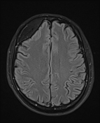
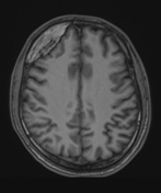
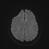
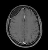
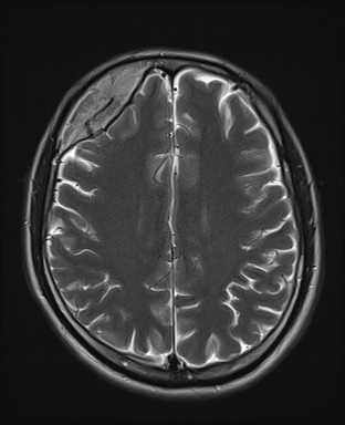
A well-defined expansive lesion is observed in the right frontal bone with hyperintense signal on T1 and T2 weighted images and hypointense signal on fat-suppressed images. There is no enhancement. The lesion compresses the frontal lobe, without edema.
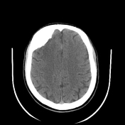


A well-defined expansive lesion is present in the right frontal bone, exhibiting heterogeneous hyperdensity (400-1000 HU). Sclerotic bands are present in the center of the lesion. No lytic lesions are observed.
The patient underwent surgery for the complete removal of all bone lesions. A titanium skull cap was reshaped and secured in place using titanium screws.
Gross description
The specimen is a fragment of the skull bone measuring 8 x 5 x 0.8 cm. The outer surface is smooth, while the inner surface exhibits a rough area. In the center, there is a raised portion measuring 3.8 x 3.5 x 2 cm, with a cut surface showing a yellow-brown color, hardness, and firmness. The specimen was divided into nine pieces, placed in nine cassettes, and preserved.
Microscopic description
Examination of the nine pieces stained with hematoxylin and eosin reveals that the cut sections contain bone tissue. In the central region, there is a proliferative area resembling Haversian bone sheets closely arranged, with interspersed small marrow cavities, mostly fatty tissue, and minimal hematopoietic marrow. Osteoblast rims are not observed, and there are no specific signs of inflammatory or malignant lesions.
Conclusion
Case Discussion
Our initial diagnosis was a calvarial osteoma, and the differential diagnosis included intraosseous meningioma, fibrous dysplasia, or even a hemangioma.
The patient underwent removal surgery, confirming the osteoma diagnosis.
Unlike more common compact osteomas, cancellous (or spongious) osteomas may contain central marrow, explaining the hyperintense signal on T1, T2, and hypointense signal on fat-suppressed images, indicating fat-containing marrow.


 Unable to process the form. Check for errors and try again.