
Presentation
Dyspnea after coronary catheterization.
Patient Data
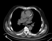

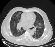
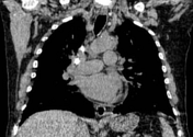
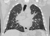

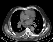
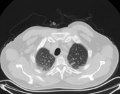
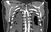
Small bilateral patchy areas of ground glass opacities are seen in both lungs with a central peribronchovascular distribution, associated with smooth subpleural and interlobular septal thickening (which indicates interstitial edema) and bilateral small pleural effusion.
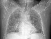
A hyperdense stent is seen within the left anterior descending artery (LAD).
Reactive mediastinal lymphadenopathy is also seen.
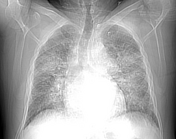
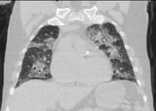

Follow-up after 2 days revealed larger areas of bilateral airspace opacification with a central peribronchovascular distribution (giving batwing appearance), associated with smooth interlobular septal thickening (giving crazy paving pattern).
No change regarding the previously noted bilateral mild pleural effusion and reactive mediastinal lymphadenopathy.
Case Discussion
The patient presented with acute chest pain and was diagnosed clinically with the acute coronary syndrome. He underwent an urgent coronary catheterization & a stent was inserted into the LAD.
One day later the patient complained of dyspnea which worsened two days later.
CT findings were typical of cardiogenic pulmonary edema.


 Unable to process the form. Check for errors and try again.