
Presentation
A 45-year-old patient with ultrasound (not available) showing numerous tortuous vessels occupying the portal vein bed referred for further CT correlation. The patient has no history of hepatic disease or laboratory evidence of hepatic impairment.
Patient Data
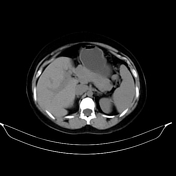

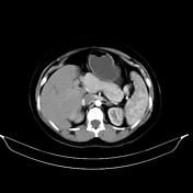

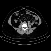
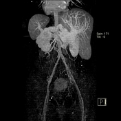

Numerous tortuous vascular channels are seen in the region of the portal ve
enhance during the portal venous phase with dilated tortuous splenic vein an
superior mesenteric vein. No filling of contrast within the described vascular channels on arterial phase. No intra-luminal thrombosis seen.These vessels drain into the left and right portal veins with additional communications with the pericholecystic veins.
Non-cirrhotic liver showing enlarged segment IV of the left lobe and reduced size of segments II and III. No hepatic focal lesions.No biliary radicles dilatation.
The spleen is not enlarged.
Case Discussion
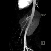
The presence of numerous vascular structures in the region of the portal vein, which enhance during the portal venous phase is diagnostic of the Cavernous transformation of the portal vein.
The lack of contrast filling during the arterial phase distinguish it from an arteriovenous malformation.


 Unable to process the form. Check for errors and try again.