
Presentation
Upper gastrointestinal bleeding secondary to esophageal varices
Patient Data
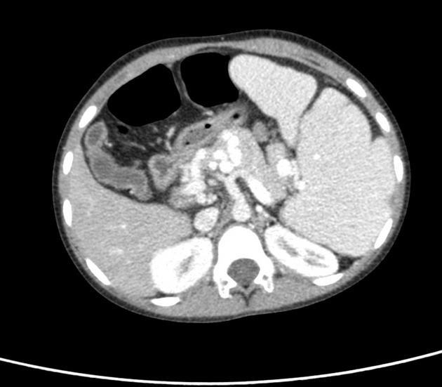
The main portal vein is not demonstrated. Instead, multiple serpentine enhancing collateral vessels are seen in the peripancreatic region and porta hepatis indicative of cavernous transformation of the portal vein. Draining into the collateral vessels are the dilated splenic and left gastric veins. The superior mesenteric vein and its tributaries are likewise dilated and appear to be drained by the dilated pancreaticoduodenal vein which eventually drains into the aforementioned collaterals. Paraesophageal, esophageal, and gastric varices are demonstrated. The non-dilated inferior mesenteric vein gradually tapers near the pancreas and eventually drains into the small caliber collaterals. The right and left intrahepatic branches of the portal vein are attenuated, more severe in the left.
The liver shows atrophic left lobe with hypertrophy of the caudate lobe.
The spleen is markedly enlarged (splenomegaly).
Case Discussion
This case demonstrates the typical appearances of cavernous transformation of the portal vein in a pediatric patient with chronic portal vein occlusion.


 Unable to process the form. Check for errors and try again.