
Presentation
Severe headache.
Patient Data
Age: 30 years
Gender: Male
From the case:
Cavum septum pellucidum and vergae


Download
Info
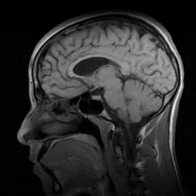
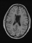
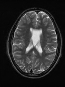
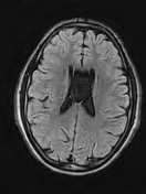
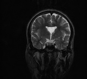
Incidental CSF containing space is seen between the frontal horns and body of the lateral ventricles representing cavum septum pellucidum associated with cavum verge (normal variant) otherwise normal brain.
left maxillary sinus retension cyst.
Case Discussion
Features are typical of a cavum septum pellucidum and associated cavum vergae.


 Unable to process the form. Check for errors and try again.