
Presentation
History of breast cancer 2 years ago currently presents with headache, left eye blindness and recent onset of left ocular swelling. Known brain metastasis before left eye swelling.
Patient Data
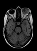

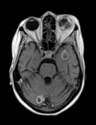
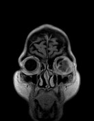
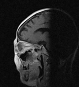

Multiple scattered bilateral focal lesions are noted within both cerebral and cerebellar hemispheres, the largest is reaching 23 mm at the right high parietal region. They showed peripheral ring enhancement sparing central non-enhancing breaking down of fluid like intensities. Mild perilesional oedema could be noted.
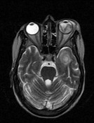
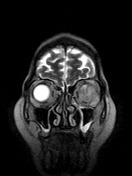
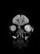
The left orbit showed intra and extra-ocular space occupying lesion related to the lateral and inferior sides of the eye globe indenting the vitreous and extending along the anterior and inferior aspects of the eye globe towards left lower eyelid. It is measuring up to 25 x 30 x 30 mm in AP, SS and CC dimensions respectively. It also showed peripheral enhancement and central breaking down as well.
Case Discussion
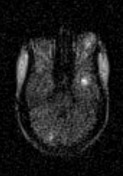
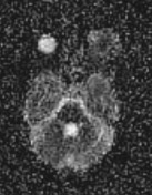
In the context of a history of breast cancer 2 years ago, established brain metastasis was considered and the recently identified left ocular lesion is likely to be ocular metastasis (uveal metastasis). It showed the same radiological features of the brain parenchymal lesion as the marginal enhancement and central necrosis. The possibility of being a primary tumour is less likely as for example the primary uveal malignant melanoma is usually hyperintense on T1, hypointense on T2 with more solid enhancement on T1C+. Also the presence of the brain metastatic lesions before the ocular lesion. It showed restriction on DWI as shown by some brain parenchymal lesions.


 Unable to process the form. Check for errors and try again.