
Cerebellar, hippocampal, and basal nuclei transient oedema with restricted diffusion (CHANTER) syndrome
Presentation
Patient with a history of polysubstance abuse is brought in by emergency medical services obtunded. Urine toxicology is positive for fentanyl.
Patient Data
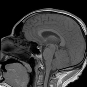

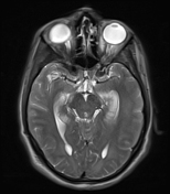
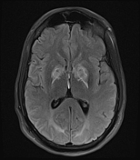
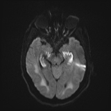
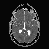
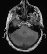


Contrast enhanced MRI of the brain displays symmetric T2/FLAIR hyperintensity throughout the bilateral cerebellar hemispheres, hippocampi, and basal ganglia, as well as diffusely throughout the cerebral white matter. T2/FLAIR hyperintensity is associated with diffusion restriction and swelling. There is significant mass effect within the posterior fossa evidenced by partial effacement of the 4th ventricle as well as the prepontine and quadrigeminal cisterns.
Following contrast administration, there is patchy parenchymal enhancement most notable in the cerebellar hemispheres.
Case Discussion
This case represents an extensive yet somewhat atypical appearance of CHANTER syndrome. The symmetric involvement of the cerebellar hemispheres, hippocampi, and basal ganglia (globus pallidi in particular) is classic. However, diffuse cerebral white matter involvement is not common with CHANTER syndrome and is more often seen in heroin-induced leukoencephalopathy.


 Unable to process the form. Check for errors and try again.