
Presentation
Three-month history of generalised headache and ataxia.
Patient Data
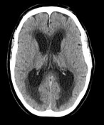

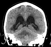
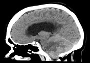
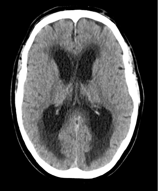
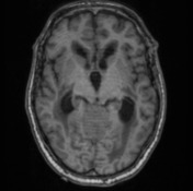
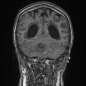
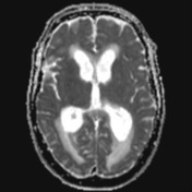
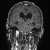
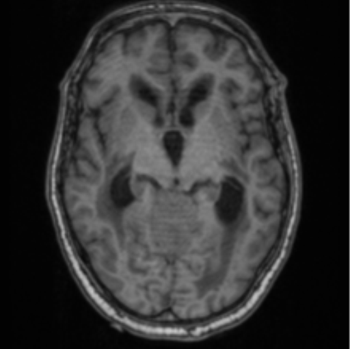
Moderate dilatation of both lateral ventricles with extensive low attenuation of the surrounding white matter consistent with transependymal oedema. Marked heterogeneity of the cerebellar hemispheres more prominent on the right side and distortion of the fourth ventricle. Early tonsillar herniation no more than 5mm in depth.
No intracranial haemorrhage of infarction.
Conclusion: Poorly defined right cerebellar lesion likely metastatic disease causing severe mass effect and obstructive hydrocephalus.
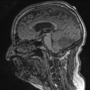
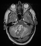
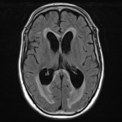
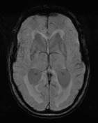
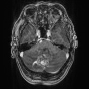
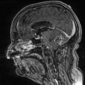
Right cerebellar mass extending into the left side with likely involvement of the vermis. Central high T2 signal hyperintensity and T2/FLAIR heterogeneity is demonstrated. Peripheral enhancement with corresponding abnormally low ADC value.
Mass effect on adjacent structures with effacement of the fourth ventricle. Enlarged lateral ventricles and third ventricle with surrounding T2/FLAIR hyperintensity within the periventricular white matter is consistent with hydocephalus and associated transependymal oedema.
Conclusion: Right cerebellar mass with obstructive hydrocephalus. Differentials include metastatic disease and high grade glioma.
Case Discussion
The patient underwent stereotactic posterior fossa craniotomy and gross total resection of the right cerebellar lesion. He had a Medical background of recently diagnosed lung adenocarcinoma.
HISTOLOGY:
Sections show extensively necrotic tumour forming sheets and large nests of cohesive epithelioid cells with large hyperchromatic pleomorphic nuclei, clumped chromatin, occasional prominent nucleoli and moderate amount of vacuolated cytoplasm. No definite squamous or glandular differentiation.
- Positive
- AE1/3
- CK7
- TTF1
- Negative
- CK20
- p40
- MelanA
- SOX10
FINAL DIAGNOSIS: Metastatic non-small cell carcinoma, favouring lung adenocarcinoma.


 Unable to process the form. Check for errors and try again.