
Presentation
Headache and ataxia.
Patient Data


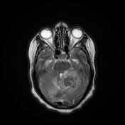
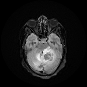
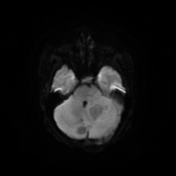
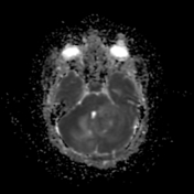
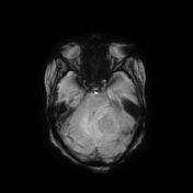
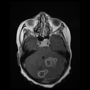
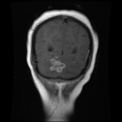
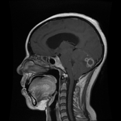
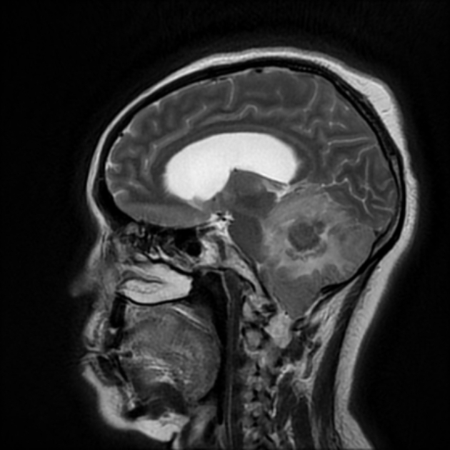
Bilateral cerebellar space-occupying lesions eliciting low signal on T2 and FLAIR WI. They appear as coalescent ring-enhancing nodules merging into large lesions. The lesions are surrounded by vasogenic edema signal and exert positive mass effect in the form of compression of the 4th ventricle and subsequent moderate supratentorial hydrocephalus, as well as mild cerebellar tonsillar herniation.
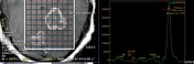
MRS showed a markedly elevated lipid/lactate peak. No significant elevation of the Choline peak.
Radiological findings with the characteristic low T2 signal and coalescent ring-enhancing nodules are suggestive of cerebellar tuberculomas.
The patient tested positive for sputum acid-fast bacillus (AFB) and Genexpert for tuberculosis.
CSF analysis revealed high protein, low glucose, and was positive for AFB.
A CT chest was performed to detect pulmonary tuberculosis.
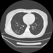
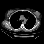

Bilateral lower lobar tree-in-bud infiltrates, more on the right. No pulmonary consolidation or cavitation.
Case Discussion
The coalescent ring-enhancing lesions with characteristic low T2W/FLAIR signal raised suspicion of granulomatous disease, likely tuberculosis.
Tuberculosis of the central nervous system can result from either hematogenous spread from distant systemic infection (e.g. pulmonary tuberculosis), as in our case.


 Unable to process the form. Check for errors and try again.