
Presentation
Acute altered state of consciousness and seizure.
Patient Data

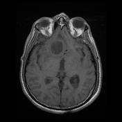
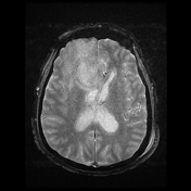
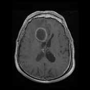

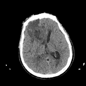
oval-shaped intra-axial peripherally-enhancing lesion located in the right frontal lobe associated with a significant amount of surrounding vasogenic edema
mass effect with an 8 mm right-to-left midline shift with subfalcine herniation
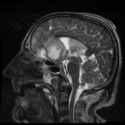
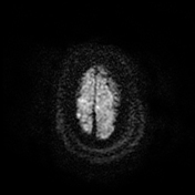
complete and smooth T2* hypointense rim overlapping with the contrast-enhancing rim
two concentric rims surround the abscess cavity, the outer one is hypointense, and the inner one is more hyperintense: dual rim sign
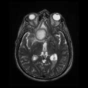
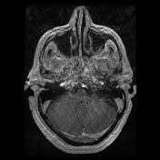
thickening of the ependyma of the lateral ventricles, layering debris with a fluid-fluid level in both occipital horns in keeping with ventriculitis
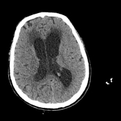
non-communicating hydrocephalus with dilatation of the lateral ventricles and the third ventricle
bilateral enhancement of the choroid plexus in keeping with choroid plexitis
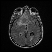
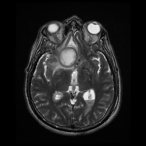
left temporal enhancement around the left M1 segment which demostrates parietal irregularities in keeping with vasculoapthy
thichkening and enhcancement of the
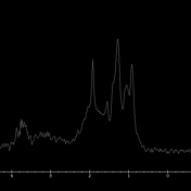
no choline peak nor NAA decrease on MRS
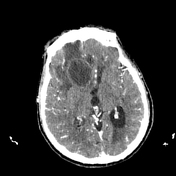

oval-shaped intra-axial peripherally-enhancing lesion located in the right frontal lobe
surrounding vasogenic edema and mass effect with subflacine herniation.
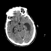
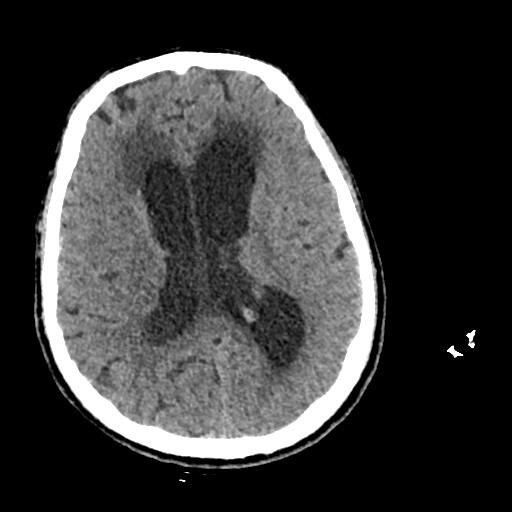
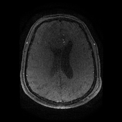
Post-operative CT demonstrates the regression of the mass effect and the subflacine herniation after the surgical drainage of the abscess.
Case Discussion
Features of a cerebral abscess complicated with ventriculitis, hydrocephalus, choroid plexitis, ischemic infarcts, and vasculopathy.


 Unable to process the form. Check for errors and try again.