
Presentation
Left hemiparesis, diplopia, loss of balance, and vomiting.
Patient Data


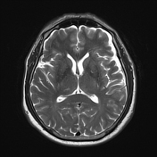
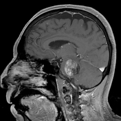

At the level of the pons, an intra-axial lesion with irregular morphology, defined lobulated borders, heterogeneous appearance, predominantly hyperintense on T1, hypointense on T2 and FLAIR, without diffusion restriction (not shown).
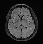
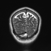
Multiple other smaller similar lesions are seen elsewhere.
Incidental left frontal developmental venous anomaly.
Case Discussion
This patient had a history of an ulcer on the left foot that progressively increased in size and was diagnosed as nodular melanoma based on histopathological results. This type represents approximately 15% of melanomas. It often appears quickly as a bump on the skin. It is usually black but can be pink or red.
Melanoma is the third most common cause of brain metastases, after lung cancer and breast. It can affect the brain and leptomeninges. In 75% of cases, metastatic lesions are multiple.


 Unable to process the form. Check for errors and try again.