
Presentation
Neck pain
Patient Data
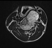

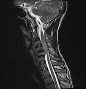
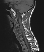
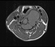
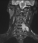
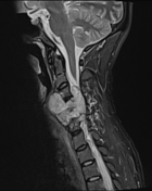
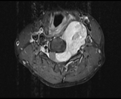
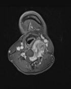

A well-defined lobulated enhancing solid mass lesion in the extramedullary intradural space in the spinal canal on the left side, extending from C3 to C6 level. It extends into paravertebral space through C3-C4, C4-C5 & C5-С6 widened intervertebral foramina on the left side, giving a dumbbell-shaped appearance. Large para vertebral component seen causing posterior displacement of vertebral artery with encasement of the vertebral artery at the place, however, flow void is maintained and no luminal narrowing seen. Marked spinal cord compression at these levels by the above mass lesion with right lateral deviation of the cord and intramedullary T2 W hyperintense cord signal abnormality at C3 & C4 levels.
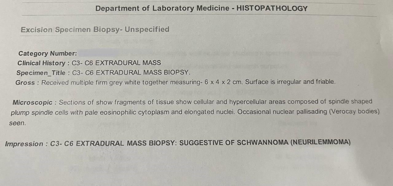
The histopathological report suggestive of schwannoma (neurilemmoma).
Case Discussion
Spinal schwannoma is benign nerve sheath tumors within the spinal canal, typically arising from spinal nerve roots and it is the most common nerve sheath tumor of spine.
Co-author: Dr. Mukund Prasad (M.ch Neurosurgery).


 Unable to process the form. Check for errors and try again.