
Presentation
G1P0 at 22 weeks gestation. Abnormal ultrasound exam. MRI requested by her gynaecologist.
Patient Data
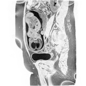

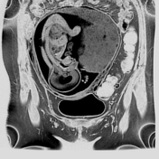

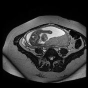
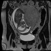

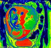
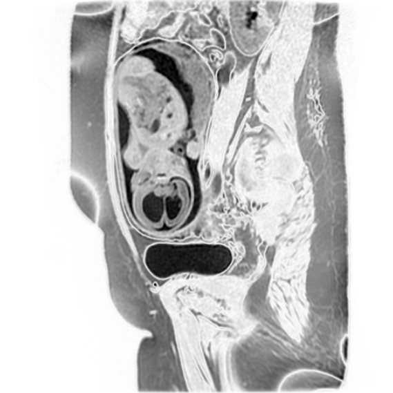
Low lying cerebellar tonsils which are extended below the foramen magnum with laminated 4th ventricle, and dilated supratentorial ventricular system (3rd, and lateral ventricles).
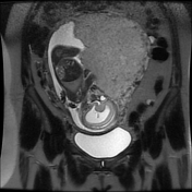
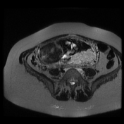
There is a midline cystic mass with no skin cover at the lower dorsal region, communicating with the spinal canal through a spinal dysraphism (spina bifida aperta). The adjacent segment of the spinal cord is angulated towards the cystic mass, indicating myelomeningocele.
No other associated fetal malformations were seen.
Normal amount of amniotic fluid.
Placenta corporeal/fundal posterior 6.5 cm from the internal os.
Case Discussion
MRI features of Chiari II malformation, also known as Arnold-Chiari malformation.
MRI is superior to ultrasound in the analysis of the cerebral posterior fossa as well as in the detection of associated fetal malformations.


 Unable to process the form. Check for errors and try again.