
Presentation
Severe epigastric pain.
Patient Data
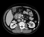

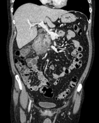

The first and second parts of the duodenum shows oedematous wall with surrounding inflammatory changes seen as stranding of fat planes, multiple small encysted paraduodenal collections and thickening of right Gerota fascia, suggestive of paraduodenal pancreatitis.
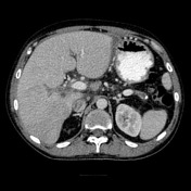
The pancreatic head shows multiple hypodense areas with coarse calcifications posteriorly, suggestive of dilated pancreatic ducts with stone formation.
Dilated entire main pancreatic duct with multiple calculi and atrophic changes at pancreatic tail.
Hepatic pseudolesion near falciform ligament is incidentally noted.
The patient has history of recurrent abdominal pain, the last attack was since one year.
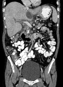

Mild decreased parenchymal enhancement of the pancreas with peripancreatic fat stranding, right Gerota fascia thickening, and thickened wall of the first and second parts of the duodenum.
Dilated entire pancreatic duct with multiple calculi, and coarse calcification at the posterior part of pancreatic head likely calculi.
Floating thrombus is seen at the left main portal vein.
No enhancement of right main portal vein and its branches, suggestive of acute thrombosis.
Diffuse interstitial oedema of the liver. Two caudate lobe parenchymal cystic lesions are seen. A subhepatic collection at the free edge of left lobe.
Multiple portocaval, peripancreatic and upper abdominal reactive lymph nodes are noted.
Case Discussion
Two episodes of acute pancreatitis on top of chronic pancreatitis in 1 year interval.
The main signs of chronic pancreatitis are:
dilated main pancreatic duct with multiple calculi
atrophic changes of the pancreas with parenchymal calcifications
pancreatic pseudocysts


 Unable to process the form. Check for errors and try again.