
Presentation
Chronic cough and shortness of breath.
Patient Data
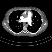


There are wall thickening and internal bands in the lower lobar and segmental branches of the bilateral pulmonary arteries. The upper lobar and segmental branches on both sides also exhibit internal bands with focal strictures. These findings are indicative of chronic pulmonary thromboembolism.
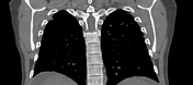
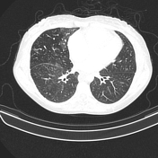
There is consequent mosaic perfusion of both lungs due to chronic thromboembolism.
Bilateral lung subpleural atelectatic bands and a few small patches suggest possible small lung infarcts.
Increased diameter of the pulmonary trunk compared to the adjacent ascending aorta and dilated right ventricle suggests pulmonary hypertension.
There is a reflux of contrast from the right atrium into the hepatic segment of the inferior vena cava during the early arterial phase, suggesting right side heart impairment.
The upper abdominal cuts show cirrhotic liver changes with moderate ascites.
Case Discussion
This case shows features of bilateral pulmonary chronic thromboembolism with consequent pulmonary hypertension and mosaic perfusion of both lungs.
Chronic pulmonary emboli are mainly a consequence of incomplete resolution of pulmonary thromboembolism.
Pulmonary chronic thromboembolism is on of the causes of mosaic attenuation of the lung.


 Unable to process the form. Check for errors and try again.