
Presentation
Right flank pain. No nausea, vomiting or urinary symptoms.
Patient Data
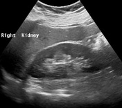
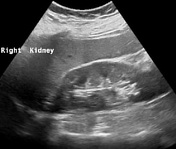
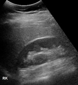
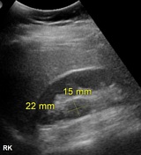

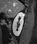
Well-defined isoechoic lesion measuring 15 x 22 mm, at the upper pole of right kidney. No renal calculi or hydronephrosis is seen.


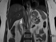
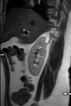
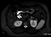
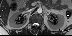
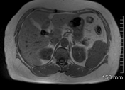
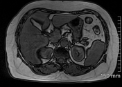

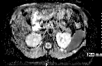
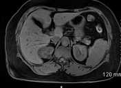
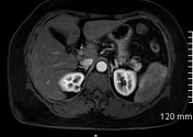
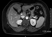
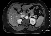
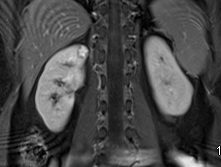
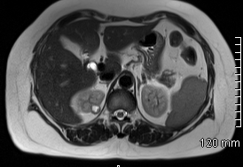
Small well-defined exophytic lesion measuring about 15 x 21 mm arising from the posterior upper pole of the right kidney. It is isointense on T1, hyperintense on T2-weighted images and heterogeneously enhance on the post contrast study. It shows no diffusion restriction. No fat, calcifications or hemorrhage is seen in it. The right renal vein and IVC are patent. No perinephric infiltration is noted. No loco-regional lymphadenopathy is seen. Adrenals are normal. Tiny simple cyst at the upper pole of the left kidney. Status post cholecystectomy.

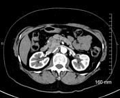
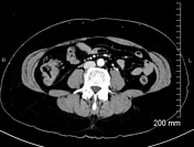
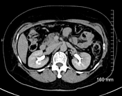

Kidneys are normal in size, shape and parenchymal density/enhancement. Redemonstration of a small exophytic lesion at the upper posterior pole of the right kidney. No calcifications or fat density is seen in it. The lesion shows peripheral enhancement on the arterial phase with progressive filling in during the venous and delayed phases. Renal veins and IVC are patent. No evidence of loco-regional or distant metastases is noted. Status post cholecystectomy.
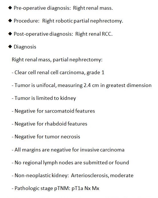
Histopathology report of the robotic right partial nephrectomy showing grade 1 clear cell renal cell carcinoma.
Case Discussion
Small well-defined exophytic lesion at the upper pole of the right kidney, showing no significant interval growth on the MRI and CT scan when compared with the baseline ultrasound examination. The lesion is suspicious for low-grade malignancy. The patient underwent an uneventful robotic partial nephrectomy and histopathology showed grade 1 clear cell renal cell carcinoma.


 Unable to process the form. Check for errors and try again.