
Presentation
Acute onset of abdominal pain in right abdomen
Patient Data
Age: 40 years
Gender: Male
From the case:
Colostomy stenosis


Download
Info
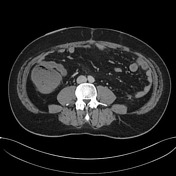
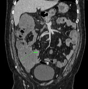
- pathologic dilatation of caecum and ascending colon. Proximal to the ileocaecal valve small bowel present a normal calibre. In the right flank, a stenosis from the ostomy is noticed.
- Hartmann pouch in pelvis. Sigmoid, descending, and transverse colon are missing.
- atrophy of the pancreas body and tail is observed due to pancreatitis sequels.
From the case:
Colostomy stenosis


Download
Info
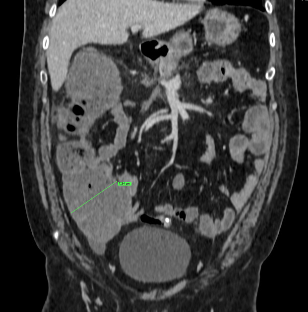
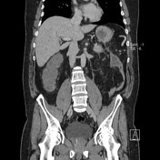
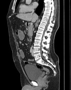
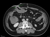
- caecum is dilated measuring 7.3 cm
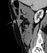
- there is a calibre change at the colostomy site
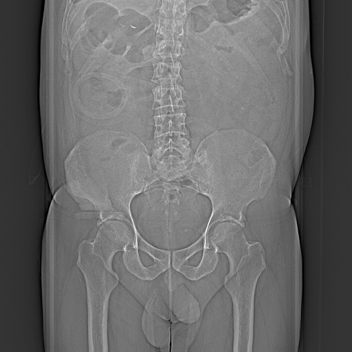
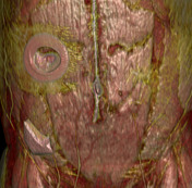
- 3D volumetric-reconstruction of the abdominal wall is displayed
- photo of colostomy
Case Discussion
The main cause of stomal stenosis is ischaemia. However, local infections, stomal retraction, and an inadequate skin opening may also lead to stenosis. Stomal stenosis may develop immediately after surgery or months later. The reported incidence varies from 2 to 14%. The majority of cases can be treated conservatively by altering the diet as long as there are no major cutaneous complications.


 Unable to process the form. Check for errors and try again.