Presentation
Recent upper endoscopy with worsening abdominal pain.
Patient Data


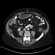
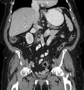
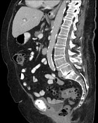
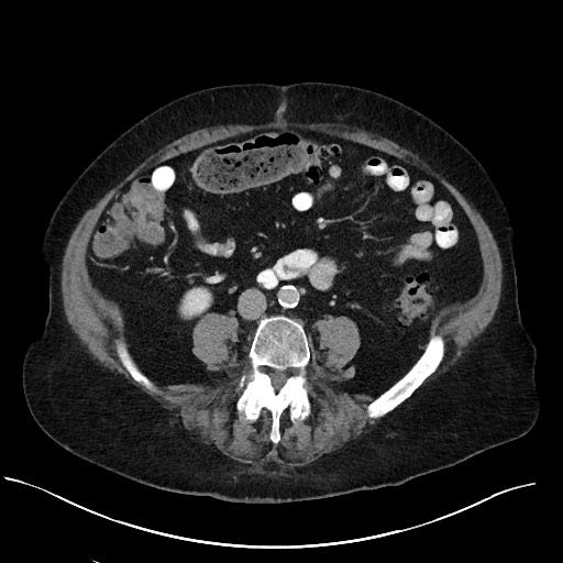
Somewhat linear, strandy intermediate-high density filling defect in the distal common bile duct. This is best appreciated on axial and coronal images. Heterogeneous areas of low attenuation within the right hepatic lobe, likely related to inflammation following ERCP or geographic fat. Mild pneumobilia. Incidental duodenal diverticulum filled with oral contrast projecting from the posterior 2nd segment.
Case Discussion
This patient presented with worsening abdominal pain following upper endoscopy. There are two findings to account for this patient's pain. The first is heterogeneous areas of hypoenhancement throughout the right hepatic lobe, which could represent inflammation from cholangitis following retrograde injection of contrast (alternatively, this could be related to heterogeneous perfusion or geographic fat).
The second finding is a somewhat linear, intermediate to high attenuation structure within the distal common bile duct. Given that this patient underwent sphincterotomy, this likely represents blood products/thrombus within the distal common bile duct. Because the ordering provider did not feel that this is resulting in significant obstruction based on the liver function tests, no further intervention was performed.


 Unable to process the form. Check for errors and try again.